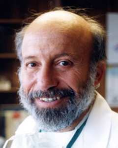
Sherman SILBER (USA)
For over 38 years Dr. Silber has originally developed all of the most popular fertility treatments used all around the world today.
He performed the world’s first microsurgical vasectomy reversal, as well as the first testicle transplant, in the 70’s and now in the current century, the world’s first ovary transplant. He was the first to develop the TESE and MESA techniques for retrieving testicular and epididymal sperm in azoospermic men. He headed the clinical MIT team that first mapped and sequenced the Y chromosome in infertile men and discovered the now famous DAZ gene for male fertility. His research includes also the study of reproduction and fertility in zoo animals and endangered species. Most recently he has perfected the preservation of fertility for cancer patients with ovarian freezing and transplantation and thereby figured out how to extend the reproductive biological clock of women.
Dr. Silber went to medical school at the University of Michigan, did post-graduate training at Stanford University, and then again at the University of Michigan. From 1967 to 1969, he provided medical care via the U. S. Public Health Service to Eskimos, Indians, and Aleuts. Then he taught at the University of Melbourne Medical School in Australia, and later at the University of California Medical School in San Francisco. He is a scientific collaborator at MIT in Cambridge, Massachusetts, at the Kato Clinic in Tokyo, and is a full professor at the University of Amsterdam, and at Sun Yat Sen University Medical School in China. His major clinical medical practice is at St. Luke’s Hospital in St. Louis, Missouri.
Abstract
The Aged Ovary
The first question to ask is, what is the intrinsic fecundity rate of the human oocyte in relation to age? Ovarian hyperstimulation has been found to yield a live baby rate per oocyte of only about 4 to 6%. Thus on average more than 20 to 25 human oocytes would be required to produce a single live baby. But that is in stimulated cycles. The live baby rate per egg in natural cycle, single embryo transfer IVF, would estimate better the intrinsic fertility of the single human oocyte in relation to age. The primary data of live baby per oocyte were approximated with a logistic curve r+1(a+exp[b(t-c)]) where r is live baby rate per oocyte and t is age in years. The coefficients were evaluated using gradient method as implemented in statistical package R (version 3.2.5). This allowed the construction of a robust logistic curve fit to relate the natural fertility of a single human oocyte to its age.
The drop in intrinsic fertility per oocyte is summarized remarkably robustly in this logistic curve. There is at first a steady (almost horizontal) maintenance of fertility per oocyte (26%) during the twenties and early thirties, followed after age 34 with a sharp linear decline of 10% per year until age 42 (4%). This decline slows down after age 43, with only 3% of original fertility remaining at age 45. In Cobo’s COH study with ovarian stimulation the live baby rate per oocyte was only 6.6% for women under 35. For Patrizio’s COH study the live baby rate per oocyte was 6.5% for women under 37. However, for natural cycle and minimal stimulation IVF in women under age 35 the live baby rate per oocyte is 26%. This is five times greater live baby rate per egg than has been reported with stimulated cycles, and is more reflective of the true natural decline in fertility of the ovary as it ages.
However this decline in oocyte fertility with age is separate from the decline in ovarian reserve (oocyte number) with age. There is also a steady decline in the number of oocytes per ovary from 6 million at 20 weeks fetal life to 2 million at birth and about 400,000 in the teen years. On average the ovary runs out of eggs by age 51, the natural almost universal mean age of menopause in most cultures, ethnic, and racial groups. What controls this decline in oocyte number, and can we delay this oocyte loss and delay menopause?
Evidence for the mechanism of this decline with age in numbers of oocytes in the human comes from experience with ovary cryopreservation and transplantation. Worldwide there have been over 148 documented and peer reviewed live births from frozen ovary tissue. The majority have been case reports. Nonetheless based on the tabulated series of young patients (i.e under 35 years old when frozen) in Brussels, Israel, Denmark, and our center in St. Louis, live birth rates vary from 41% to 76%, and most are spontaneous pregnancies not requiring IVF. But these were young women (under age 35) who had ovary cortex frozen prior to gonadotoxic treatment for cancer or other diseases. For women whose ovary cortex was frozen at an older age the success rate would be considerably lower. Nonetheless ovary tissue cryopreservation, at least for young cancer patients, is a relatively robust method for preserving a young woman’s fertility until she may be many decades older.
The mechanism for slowing down the follicle depletion of aging comes from observing in detail what happens from transplanting young frozen ovary tissue back to women when they are many years older. Patients almost always have return of ovarian function 5 months post transplant with regular menstrual cycling. AMH rises to very high levels at the same time that the FSH declines to normal. Four to eight months after the FSH returns to normal, the AMH declines again to very low levels indicating severe follicle depletion. Nonetheless, these ovary grafts remain functional for up to 5 years or longer despite very low AMH levels. Small pieces of young ovarian tissue with very low AMH levels last a very long time. These findings are surprising at first and intriguing. We can gain scientific insight from these results of ovary transplantation, into the seemingly mysterious mechanism of primordial follicle recruitment. Cortical tissue pressure seems to be the key regulator of oocyte arrest in the fetus, as well as primordial follicle recruitment in the adult, and indeed ovarian longevity.
Even for healthy women just wanting to put off child bearing, a simple ovary freeze would cost less than 6,000 dollars, and require just one simple procedure and no hormonal stimulation. Interestingly it is known from huge population studies that unilateral oophorectomy does not cause menopause to occur more than just a few years earlier. This is because as the ovarian reserve goes down, the rate of primordial follicle recruitment also goes down. Furthermore, with very few eggs, a young ovary can be quite fertile. Transplanting back ovarian tissue can give back hormonal function even after these woman might otherwise be in menopause 20 years later. And since a small piece of ovarian tissue can last a very long time, theoretically, Anderson has shown that one can theoretically continue to transplant small pieces of ovarian cortical tissue until they are even 100 years old and never go into menopause. Many female business executives and physicians have chosen this approach, which is otherwise less popular than oocyte freezing. For young women with cancer it is clearly the best approach. Of most interest, it gives an insight into the regulation of primordial follicle recruitment and longevity in the aged ovary by cortical tissue pressure and remaining ovarian reserve.
Increasing atmospheric pressure in the incubator with IPS cell derived oocytes will arrest their development and form primordial follicles which “lock” the immature oocyte and drastically retard their recruitment and development, because the gene FOX3 is made to go intra-nuclear. In contrast, dissolving the fibrous tissue, which decreases tissue pressure, causes Fox3 to go extranuclear, and thus releases the “locked” primordial follicles. Thus cortical tissue pressure is found to be a key regulator of fetal primordial follicle arrest, adult primordial follicle recruitment, and ovarian longevity. In addition, the long duration of function of such small pieces of ovarian tissue with very low AMH levels supports the postulate that when there is low ovarian reserve, the rate of primordial follicle recruitment is reduced, promoting ovarian longevity even as the follicle count is going down. This explains why in POF (premature ovarian failure) there are often still functional follicles that can be recruited with fragmentation and in vitro activation. The over recruitment of primordial follicles after transplant is consistent with tissue pressure being a. key regulator of primordial follicle recruitment and ovarian longevity.
Sudden loss of tissue pressure after transplant results in massive primordial follicle recruitment. With healing, the return of cortical tissue pressure with a high “fibrous tissue to follicle” ratio then reduces the rate of primordial follicle recruitment. As the ovarian cortex becomes depleted of primordial follicles, there is greater tissue pressure, thus slowing the rate of primordial follicle recruitment. Increasing pressure during oocyte culture will therefore arrest their development and form primordial follicles. In contrast, dissolving the fibrous tissue, which decreases tissue pressure, causes Fox3 to go extranuclear, and thus releases the “locked” primordial follicles.
Cortical tissue pressure is thus a key factor in preserving ovarian reserve in terms of just the number of eggs and the age of menopause. In addition, the long duration of function of such small pieces of ovarian tissue with very low AMH levels supports the postulate that when there is low ovarian reserve, the rate of primordial follicle recruitment is reduced, promoting ovarian longevity even as the follicle count is going down. But the aged ovary, even if still functioning because of lower primordial follicle recruitment rate, still has oocytes that have a very low “intrinsic fertility”. That is why young ovarian tissue (frozen in time) can last a very long time when transplanted back, and result in older women not only delaying menopause, but allow her at a much older age to be able to spontaneously conceive without IVF, and thus retain her fertility as well as avoid menopause.
